Hauptinhalt
Topinformationen
Research
The central theme of our research adresses functional insulator-supported nanosystems – namely molecular and metal-coordination structures on mineral surfaces – for sustainable future technology. Insulator mineral surfaces are a central topic as this environment offers the preservation of instrinsic molecular properties via electronic decoupling.
We develop and characterise materials that offer the potential to realise field-coupled nanocomputing concepts such as the molecular quantum-dot cellular automaton (mQCA) or the development of molecular neuromorphic computing architectures.
Our research can be divided into five research lines:
- (A) Structure and reactivity of mineral surfaces
- (B) Functional nanostructures on insulator surfaces
- (C) Measurement and control of single elementary charges
- (D) Atomic-force microscopy for high-precision measurements
- (E) Method development
— Structure and reactivity of mineral surfaces
Understanding atomic-scale details that are key to geological, biological, and technological processes including research topics with regard to the origin of life.
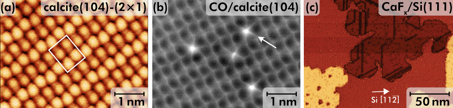
Figure 1: High-resolution imaging with non-contact atomic force microscopy of (a) the reconstructed calcite(104)-(2×1) surface and (b) single CO molecules [5]. (c) High-resolution scanning tunnelling microscopy image of a partly covered CaF1/Si(111) surface [7].
Minerals are involved in many geological, chemical, or biological processes including weathering, dissolution-precipitation, or biomineral formation. We investigate the microscopic properties of mineral surfaces with a particular interest in understanding the surface structure and chemical reactivity. Our focus currently lies on two materials classes, namely carbonates (in particular the calcite(104)-(2×1) surface) and fluorides (in particular the CaF2(111) surface).
Carbonates are among the most abundant minerals in earth’s crust and constitute a key contributor to geochemical processes. Initially, we studied the contrast formation and force interaction on calcite(104) in NC-AFM at the atomic scale using room temperature experiments [1,2,3]. We recently investigated the adsorption of carbon monoxide (CO) on this surface and confirmed the adsorption position on the calcium atoms [4]. Furthermore, we have now established that the pristine calcite(104) surface is (2×1) reconstructed, belongs to the planar space group pg, and generates two different adsorption geometries for CO, see Figure 1(a,b) [5]. Currently, we are studying the adsorption of water on this surface where we identified a particular reconstruction lifting mechanism due to water adsorption [6].
The second mineral class of interest are fluorides, in particular calcium fluoride. While this mineral is by itself a technologically most relevant material with vast application, we are particularly interested in using the (111) surface for atomic and molecular nanostructure formation. We identified advantageously similar surface properties of CaF2/CaF1/Si(111) thin films and CaF2(111) bulk crystal surfaces [7] based on STM, NC-AFM, as well as DFT data that enable the fabrication of molecular nanosystems, crossing the boundary between fundamental research and semiconductor technology. We furthermore study the interaction with scanning probe tips as a measure for the surface reactivity [8].
| [1] | P. Rahe, J. Schütte, A. Kühnle. NC-AFM contrast formation on the calcite (1014) surface. Journal of Physics: Condensed Matter, 24, 084006 (2012). |
| [2] | J. Schütte, P. Rahe, L. Tröger, S. Rode, R. Bechstein, M. Reichling, A. Kühnle. Clear signature of the (2 × 1) reconstruction of calcite(1014). Langmuir, 26, 8295 (2010). |
| [3] | S. Kuhn, M. Kittelmann, Y. Sugimoto, M. Abe, A. Kühnle, P. Rahe. Identifying the absolute orientation of a low-symmetry surface in real space. Physical Review B, 90, 195405 (2014). |
| [4] | T. M. Hafshejani, W. Wang, J. Heggemann, A. Nefedov, S. Heissler, Y. Wang, P. Rahe, P. Thissen, C. Wöll. CO adsorption on the calcite(10.4) surface: A combined experimental and theoretical study. Physical Chemistry Chemical Physics, 23, 7696 (2021). |
| [5] | J. Heggemann, Y. Ranawat, O. Krejcí, A.S. Foster, P. Rahe. Differences in molecular adsorption emanating from the (2 × 1) reconstruction of calcite(104). The Journal of Physical Chemistry Letters, 14, 1983 (2023). |
| [6] | J. Heggemann, S. Aeschlimann, T. Dickbreder, Y. S. Ranawat, R. Bechstein, A. Kühnle, A. S. Foster, and P. Rahe. Water adsorption lifts the (2 × 1) reconstruction of calcite(104). Physical Chemistry Chemical Physics, 26, 21365 (2023). |
| [7] | P. Rahe, E.F. Smith, J. Wollschläger, P.J. Moriarty. Formation routes and structural details of the CaF1 layer on Si(111) from high-resolution noncontact atomic force microscopy data. Physical Review B, 97, 125418 (2018). |
| [8] | B. Kyeyune, R. Olbrich, M. Reichling, and P. Rahe. Sublattice identification on CaF2(111): From combinatorics to physics. Physical Review B, accepted (2024). |
— Functional nanostructures
Nanomaterials for information processing past the silicon era and beyond the von-Neumann paradigm.
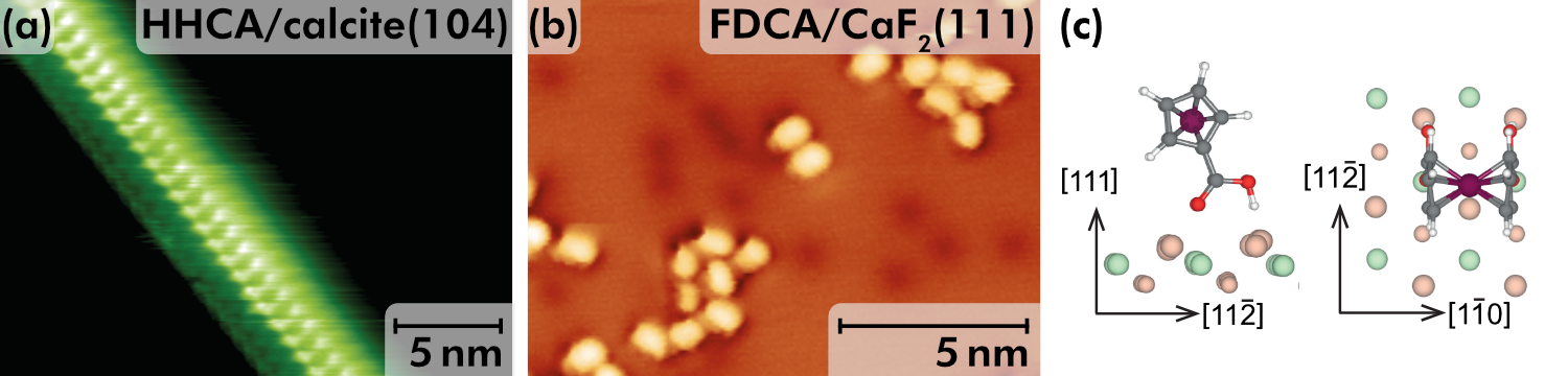
Figure 2: (a) Linear structure formed by helicene (HHCA) molecules on calcite(104) [3]; ferrocene (FDCA) molecules on CaF2(111) surfaces studied by (b) STM and (c) DFT [7].
We are interested in the fabrication, characterisation, and utilisation of functional molecule-on-insulator nanosystems to work towards novel concepts in quantum nanoscience, in particular towards novel concepts in information processing. Building upon nanoscience, chemistry, and molecular electronics, we aim for arriving at nanosystems that are build from single atoms and individual molecules, and that realise processing elements with significantly reduced energy consumption. Our focus currently lies in generating a fundamental understanding for assembly of metal-coordination materials on insulator surfaces.
We have previously studied molecular self-assembly on calcite(104) employing weak interactions such as van-der-Waals [1], hydrogen bonds [2], or combinations thereof (see Figure 2(a)) [3]. As a key result, molecular anchoring was identified as an important mechanism for molecular assembly on insulator surfaces [4]. This understanding has allowed for the realisation of on-surface synthesis on mineral surfaces [5, 6].
Currently, we investigate the formation of metal-organic structures as a key enabler for atomic-scale functionality. To name one example, we have recently characterised a quadruped binding mechanism for a functionalised ferrocene molecule on CaF2(111) (see Figure 2(b)) [7, 8] that now allows us to precisely steer the molecular structure formation. Unravelling the microscopic binding geometry for this (see Figure 2(c)) and other [9] systems is supported by density functional theory (DFT).
| [1] | P. Rahe, R. Lindner, M. Kittelmann, M. Nimmrich, A. Kühnle. From dewetting to wetting molecular layers: C60 on CaCO3(1014) as a case study. Physical Chemistry Chemical Physics, 14, 6537 (2012). |
| [2] | P. Rahe, M. Nimmrich, A. Kühnle. Substrate templating upon self-assembly of hydrogenbonded molecular networks on an insulating surface. small, 8, 2969 (2012). |
| [3] | P. Rahe, M. Nimmrich, A. Greuling, J. Schütte, I.G. Stará, J. Rybáek, G. Huerta-Angeles, I. Starý, M. Rohlfing, A. Kühnle. Toward Molecular Nanowires Self-Assembled on an Insulating Substrate: Heptahelicene-2-carboxylic acid on calcite(1014). Journal of Physical Chemistry C, 114, 1547 (2010). |
| [4] | P. Rahe, M. Kittelmann, J. L. Neff, M. Nimmrich, M. Reichling, P. Maass, A. Kühnle. Tuning molecular self-assembly on bulk insulator surfaces by anchoring of the organic building block. Advanced Materials, 25, 3948 (2013). |
| [5] | M. Kittelmann, P. Rahe, M. Nimmrich, C. M. Hauke, A. Gourdon, A. Kühnle. On-Surface Covalent Linking of Organic Building Blocks on a Bulk Insulator. ACS Nano, 5, 8420 (2011). |
| [6] | R. Lindner, P. Rahe, M. Kittelmann, A. Gourdon, R. Bechstein, A. Kühnle. Substrate Templating Guides the Photoinduced Reaction of C60 on Calcite. Angewandte Chemie, 53, 7952 (2014). |
| [7] | L. Laflör, F.A. Schlage, L. Kantorovich, P.J. Moriarty, M. Reichling, P. Rahe. Quadruped Molecular Anchoring to an Insulator: Functionalized Ferrocene on CaF2 Bulk and Thin Film Surfaces. The Journal of Physical Chemistry C, 124, 9900 (2020). |
| [8] | L. Laflör, M. Reichling, P. Rahe. Protruding hydrogen atoms as markers for the molecular orientation of a metallocene. Beilstein Journal of Nanotechnology, 11, 1432 (2020). |
| [9] | P. Rahe. PTCDA adsorption on CaF2 thin films. Beilstein Journal of Nanotechnology, 11, 1615 (2020). |
— Measurement and control of single elementary charges
Functionality following from weakly coupled to correlated electrons in nanostructures.

Figure 3: (a) Charge-state manipulation of a ferrocene island on calcite(104) [1]. (b) Central equation for quantitative charge measurements with charge force microscopy (CFM) [2, 3]. (c) Simulated CFM signal highlighting the dependency on the tip model (S, SC, SCL) and the oscillation amplitude [5].
With the functionality of nanostructures intimately bound to the behaviour of electrons within the assembly, and
with a measurement of the electronic properties allowing for a detailed characterisation of the materials, the measurement and control of single elementary charges within nanostructures is a key aspect of our research.
Previously, we pioneered charge-state modification of small molecular assemblies on calcite(104), see Figure 3(a) [1], and identified a charge stabilisation mechanism within the structure. Furthermore, we derived a consistent theory to relate the experimental measurement data to charge magnitudes with the central equation shown in Figure 3(b) [2, 3, 4, 5]. This derivation ultimately led to the introduction of charge force microscopy (CFM) [6], a technique based on the detection method of Kelvin probe force microscopy. Recently, we performed a detailed analysis of contributing factors and unravelled many influences to the absolute measurement signal [6]. In a current project, we use experimental distance-dependent data to realise quantitative charge measurements at room temperature.
Furthermore, we currently implement scanning quantum dot microscopy (SQDM) to further improve the spatial resolution and to perform quantitative measurements of electrostatic parameters at the single-molecule level. This technique uses a quantum dot attached to the tip of an atomic force microscope acting as a quantum sensor. We use molecular manipulation for the preparation of functionalised tips, in particular by attaching a single PTCDA molecule for the SQDM method.
| [1] | P. Rahe, R.P. Steele, C.C. Williams. Consecutive Charging of a Molecule-on-Insulator Ensemble Using Single Electron Tunnelling Methods. Nano Letters, 16, 911 (2016). |
| [2] | J.L. Neff, P. Rahe. Insights into Kelvin probe force microscopy data of insulator-supported molecules. Physical Review B, 91, 085424 (2015). |
| [3] | H. Söngen, P. Rahe, J.L. Neff, R. Bechstein, A. Kühnle. The weight function for charges – A rigorous theoretical concept for Kelvin probe force microscopy. Journal of Applied Physics, 119, 025304 (2016). |
| [4] | P. Rahe, H. Söngen. Imaging Static Charge Distributions: A Comprehensive KPFM Theory. In: Kelvin Probe Force Microscopy: From Single Charge Detection to Device Characterization, Eds. Sadewasser, S.; Glatzel, T., Springer Int. Publishing (2018). |
| [5] | H. Söngen, P. Rahe, R. Bechstein, A. Kühnle. Interpretation of KPFM Data with the Weight Function for Charges. In: Kelvin Probe Force Microscopy: From Single Charge Detection to Device Characterization, Eds. Sadewasser, S.; Glatzel, T., Springer Int. Publishing (2018). |
| [6] | D. Heile, R. Olbrich, M. Reichling, and P. Rahe. Modeling nanoscale charge measurements. Physical Review B, 108, 085420 (2023). |
— Atomic force microscopy for high-precision measurements
Mapping the quantum nanoscale at the frontier of spatial resolution.
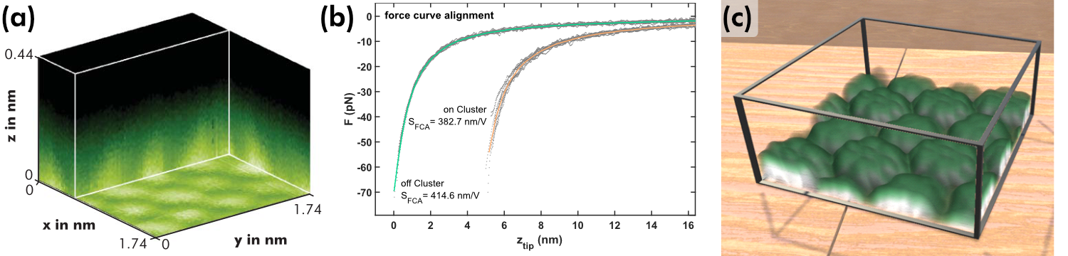
Figure 4: (a) 3D force mapping on the calcite(104) mineral surface at 300 K [12]. (b) Precise NC-AFM force measurements performed on and off single Au clusters on CeO2(111) using force curve alignment [49]. (c) 3D representation of the interaction above a PTCDA molecular island.
The core capability of frequency-modulated (also known as 'dynamic' or 'non-contact') atomic force microscopy is to quantitatively measure forces with pico-metre resolution and pico-Newton sensitivity, enabling the exciting possibility to investigate chemical or physical interactions at the atomic scale.
In the past, we applied this technique to measure the force field above surfaces such as calcite(104), see also Figure 4(a) [1], or above molecular structures such as hydrogen-bonded assemblies [2].
Furthermore, we critically analysed approaches to separate background (“long-range”) from site-specific (“short-range”) interaction forces [3] and studied the influence of an inclined tip oscillation path [4]. A recent breakthrough result is the introduction of force curve alignment (FCA) to significantly increase the precision of quantitative force measurements and to deliver an inherent consistency check, see Figure 4(b) [5].
Within the BMBF-funded project “VRnano” (https://www.vrnano.de), VR-based techniques are currently developed to allow for an efficient navigation within the three-dimensional space above a surface at the atomic scale, to manipulate single molecules by a manual “haptic” access, to visualise the many data dimensions in scanning probe microscopy with scientific rigorousness (see also Figure 4(c)), and to measure nanoscale forces between complex, three-dimensional molecules along free trajectories.
| [1] | S. Kuhn, M. Kittelmann, Y. Sugimoto, M. Abe, A. Kühnle, P. Rahe. Identifying the absolute orientation of a low-symmetry surface in real space. Physical Review B, 90, 195405 (2014). |
| [2] | A.M. Sweetman, S.P. Jarvis, H. Sang, I. Lekkas, P. Rahe, Y .Wang, J. Wang, N.R. Champness, L. Kantorovich, P.J. Moriarty. Mapping the force field of a hydrogen-bonded assembly. Nature Communications, 5, 3931 (2014). |
| [3] | S. Kuhn, P. Rahe. Discriminating short-range from van der Waals forces using total force data in noncontact atomic force microscopy. Physical Review B, 89, 235417 (2014). |
| [4] | P. Rahe, D. Heile, R. Olbrich, M. Reichling. Quantitative dynamic force microscopy with inclined tip oscillation. Beilstein Journal of Nanotechnology, 13, 610 (2022). |
| [5] | D. Heile*, R. Olbrich*, M. Reichling, P. Rahe. Alignment method for the accurate and precise quantification of tip-surface forces. Physical Review B, 103, 075409 (2021). (DH and RO contributed equally) |
— Method development
Pushing the boundaries of scanning probe microscopy.
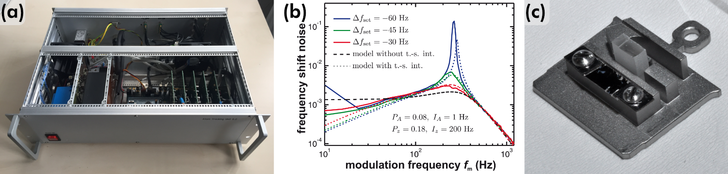
Figure 5: (a) Atom-tracking electronic unit for drift measurement and compensation [1]. (b) Measured (solid lines) and simulated (dashed lines) frequency shift noise spectrum [3]. (c) Double sample holder supporting a metallic Ag(111) and mineral calcite(104) sample [5].
The study of insulator mineral surfaces as well as molecular structures at the atomic scale in real space requires an optimised, well-characterised, and low-noise scanning probe microscope operated at the physical limits.
Thermal drift – the result of minute temperature changes within a scanning probe microscope – is a well-known nuisance, and especially hinders a reliable absolute tip positioning. It especially forbids long-term data acquisition in constant-height mode as well as the acquisition of dense force volume data. An elegant solution is to "lock" the scanning probe tip to one surface site and to track the virtual movement, a technique known as atom tracking. To measure and compensate for these deviations, we have developed an atom-tracking system, see Figure 5(a) [1]. The latest version of this system is based on an ARM Cortex-M4 microcontroller that enables force measurements over the course of many hours.
The performance of scanning probe microscopes is ultimately defined by the magnitude of measurement noise. We have previously performed a detailed analysis of NC-AFM noise figures (see Figure 5(b)), including a mathematical description of the whole detection system [2] and also including the tip-sample interaction [3].
Furthermore, we introduced the “dip-df” control mode [4] and developed tools and protocols for the efficient high-resolution study of mineral surfaces at low temperatures such as a double sample holder, see Figure 5(c) [5].
| [1] | P. Rahe, J. Schütte, W. Schniederberend, M. Reichling, M. Abe, Y. Sugimoto, A. Kühnle. Flexible drift-compensation system for precise 3D force mapping in severe drift environments. Review of Scientific Instruments, 82, 063704 (2011). |
| [2] | J. Lübbe, M. Temmen, S. Rode, P. Rahe, A. Kühnle, M. Reichling. Thermal noise limit for ultra-high vacuum non-contact atomic force microscopy. Beilstein Journal of Nanotechnology, 4, 32 (2013). |
| [3] | J. Lübbe, M. Temmen, P. Rahe, M. Reichling. Noise in NC-AFM measurements with significant tip-sample interaction. Beilstein Journal of Nanotechnology, 7, 1885 (2016). |
| [4] | S. Rode, M. Schreiber, A. Kühnle, P. Rahe. Frequency-modulated atomic force microscopy operation by imaging at the frequency shift minimum: The "dip-df" mode. Review of Scientific Instruments, 85, 043707 (2014). |
| [5] | J. Heggemann, L. Laflör, Linda, P. Rahe. Double sample holder for high-resolution studies of an insulator surface with well-defined tips. Review of Scientific Instruments, 92, 053705 (2021). |